When ultrasonic waves are used in medicine for diagnostic purposes, high-frequency sound pulses are produced by a transmitter and directed into the body. As in sonar, reflections occur. They occur each time a pulse encounters a boundary between two tissues that have different densities or a boundary between a tissue and the adjacent fluid. By scanning ultrasonic waves across the body and detecting the echoes generated from various internal locations, it is possible to obtain an image or sonogram of the inner anatomy. Ultrasonic imaging is employed extensively in obstetrics to examine the developing fetus (Figure 16.34). The fetus, surrounded by the amniotic sac, can be distinguished from other anatomical features so that fetal size, position, and possible abnormalities can be detected.
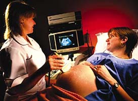 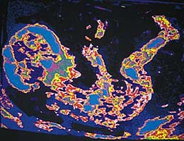 | | Figure 16.34
An ultrasonic scanner can be used to produce an image of the fetus as it develops in the uterus. (Left, © Deep Light Productions/Science Photo Library/Photo Researchers; right, © Howard Sochurek) |
|
Ultrasound is also used in other medically related areas. For instance, malignancies in the liver, kidney, brain, and pancreas can be detected with ultrasound. Yet another application involves monitoring the real-time movement of pulsating structures, such as heart valves (“echocardiography”) and large blood vessels.
When ultrasound is used to form images of internal anatomical features or foreign objects in the body, the wavelength of the sound wave must be about the same size as, or smaller than, the object to be located. Therefore, high frequencies in the range from 1 to 15 MHz (1 MHz =1 megahertz=1×10⁶ Hz) are the norm. For instance, the wavelength of 5-MHz ultrasound is l=v/f=0.3 mm, if a value of 1540 m/s is used for the speed of sound through tissue. A sound wave with a frequency higher than 5 MHz and a correspondingly shorter wavelength is required for locating objects smaller than 0.3 mm.
Ultrasound also has applications other than imaging. Neurosurgeons use a device called a cavitron ultrasonic surgical aspirator (CUSA) to remove brain tumors once thought to be inoperable. Ultrasonic sound waves cause the slender tip of the CUSA probe (see Figure 16.35) to vibrate at approximately 23 kHz. The probe shatters any section of the tumor that it touches, and the fragments are flushed out of the brain with a saline solution. Because the tip of the probe is small, the surgeon can selectively remove small bits of malignant tissue without damaging the surrounding healthy tissue.
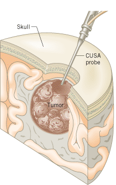 | | Figure 16.35
Neurosurgeons use a cavitron ultrasonic surgical aspirator (CUSA) to “cut out” brain tumors without adversely affecting the surrounding healthy tissue. |
|
Another application of ultrasound is in a new type of bloodless surgery, which can eliminate abnormal cells, such as those in benign hyperplasia of the prostate gland. Still in the experimental phase, this technique is known as HIFU (high-intensity focused ultrasound). It is analogous to focusing the sun’s electromagnetic waves by using a magnifying glass and producing a small region where the energy carried by the waves can cause localized heating. Ultrasonic waves can be used in a similar fashion. The waves enter directly through the skin and come into focus inside the body over a region that is sufficiently well defined to be surgically useful. Within this region the energy of the waves causes localized heating, leading to a temperature of about 56 °C (normal body temperature is 37 °C), which is sufficient to kill abnormal cells. The killed cells are eventually removed by the body’s natural processes.
The Doppler flow meter is a particularly interesting medical application of the Doppler effect. This device measures the speed of blood flow, using transmitting and receiving elements that are placed directly on the skin, as in Figure 16.36. The transmitter emits a continuous sound whose frequency is typically about 5 MHz. When the sound is reflected from the red blood cells, its frequency is changed in a kind of Doppler effect because the cells are moving. The receiving element detects the reflected sound, and an electronic counter measures its frequency, which is Doppler-shifted relative to the transmitter frequency. From the change in frequency the speed of the blood flow can be determined. Typically, the change in frequency is around 600 Hz for flow speeds of about 0.1 m/s. The Doppler flow meter can be used to locate regions where blood vessels have narrowed, since greater flow speeds occur in the narrowed regions, according to the equation of continuity (see Section 11.8). In addition, the Doppler flow meter can be used to detect the motion of a fetal heart as early as 8–10 weeks after conception.
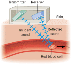 | | Figure 16.36
A Doppler flow meter measures the speed of red blood cells. |
|
|
| Copyright © 2000-2003 by John Wiley & Sons, Inc. or related companies. All rights reserved. |